Study anatomy and physiology with AI, from the chapter you’re on
A&P asks you to know every structure, what it does, and what happens when it fails. Upload your chapter and Hemle turns it into flashcards, quizzes and answers that name the page. Below is what it made from a real chapter on the heart.
The chapter we used
Chapter 19 of OpenStax’s Anatomy and Physiology, “The Cardiovascular System: The Heart”: 64 pages on the heart’s structure, its electrical system, the cardiac cycle and what sets cardiac output. It’s the book’s first edition, from 2013, which OpenStax published under a CC BY 4.0 licence. A newer edition exists, so your course’s chapter may read differently.
We uploaded the chapter as OpenStax’s PDF prints it, on 28 October 2026, and Hemle found 25 concepts in it. Everything below marked as Hemle’s is what it returned, word for word. One set of flashcards is left out, because one of its cards came back unfinished; the cards section says which.
One question, asked of the chapter
Describing the path of blood through the heart is one of the chapter’s own objectives, so we asked Hemle’s Chat to trace it. Beside the answer is page 6, one of the two pages it cites, with the passage the app marked: the caption of Figure 19.4.
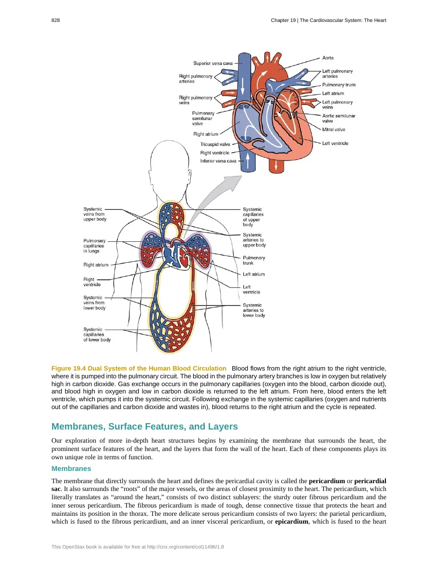
A drop of blood returning from the body enters the right atrium through the superior vena cava or inferior vena cava. It then moves to the right ventricle, which pumps it into the pulmonary trunk. The pulmonary trunk divides into the left and right pulmonary arteries, carrying the blood toward the lungs (pp. 5–6).
In the lung capillaries, carbon dioxide leaves the blood and oxygen enters it. The oxygen-rich blood returns to the heart through the pulmonary veins, enters the left atrium, and then moves into the left ventricle. The left ventricle pumps it into the aorta, which carries it out to the body. After oxygen is delivered to body tissues, the blood returns through veins to the venae cavae and the right atrium, repeating the cycle (pp. 5–6).
Does the part about blood flow through the heart make sense?
Answers are written by AI and can be wrong. Check the pages they cite.
How to study anatomy and physiology
A&P is two subjects at once: where things are, and how they work. These habits serve both, and each is shown on Chapter 19. They need only a pen and paper; where Hemle saves you time, we point to what it made.
Learn each structure with its job
Exams ask what a part does, and what goes wrong without it, as often as where it is. For each structure, write one line for where it is, one for what it does, and one for what fails without it.
In Chapter 19
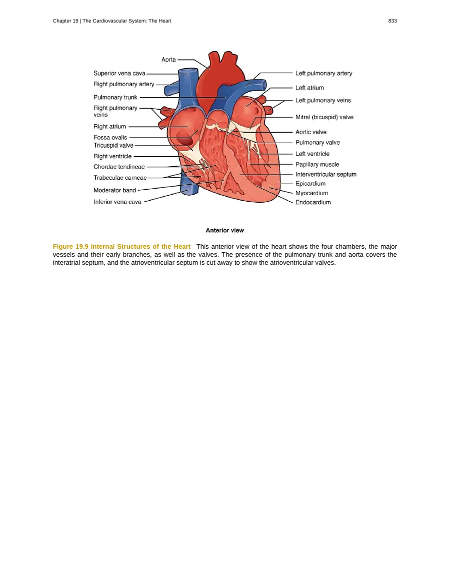
The chordae tendineae and papillary muscles hold the atrioventricular valves’ flaps in place, so they aren’t blown back into the atria when the ventricles contract (pp. 14–17).
Hemle’s cards on the heart valvesTrace pathways from memory
Blood, nerve impulses and hormones all travel routes, and exams ask for them in order. Say or draw the route with the book closed, then check it against the figure.
In Chapter 19
Two routes: blood through the chambers, valves and vessels (Figure 19.4, p. 6), and the impulse from the SA node through the AV node, the AV bundle and its branches to the Purkinje fibers (Figure 19.18, p. 26).
Hemle’s answer tracing the bloodPut what happens together on one timeline
Physiology often runs several events at once. Learn them on one shared timeline rather than as separate lists, so you can say what is happening everywhere at a given moment.
In Chapter 19
The ECG’s waves, the chambers contracting and relaxing, and the heart sounds happen together. The chapter draws them on shared time axes in Figures 19.28 and 19.29 (pp. 40–42).
Hemle’s cards on the ECG’s wavesExplain cause and effect to yourself
Physiology questions are often “what happens if…”. Explain each step in your own words: what changes, and why the next thing follows from it.
In one small study of 24 eighth graders, those who explained each sentence of a textbook unit on the circulatory system to themselves understood it better than classmates who read it twice, though they also spent longer on it. (Chi and colleagues, 1994)
In Chapter 19
Why can heart rate rise to about 100 bpm with no sympathetic stimulation at all? Because less parasympathetic stimulation lets the SA node speed up toward its own rhythm (pp. 46–47).
Hemle’s quiz on heart rate controlTake the words apart
Most A&P terms are built from a few Greek and Latin parts. Learn the common ones and a new term half explains itself.
In Chapter 19
The chapter starts with one: it traces “cardiac” back to kardia, heart (p. 2). The heart wall’s layers build on it: endocardium, myocardium, epicardium, and the pericardium around them (pp. 7–9).
Every card on four concepts
Flashcards Hemle wrote on the chapter, each set whole: the question, the answer behind it, and the pages it cites. Try each one before you open it.
ECG Waveform Components
5 cards from page 32
What electrical event does the P wave represent on an ECG, and what is its relationship to atrial contraction?Show answerHide answer
The P wave represents the depolarization of the atria. Atrial contraction begins approximately 25 ms after the start of the P wave.
Why is the QRS complex significantly larger than the P wave on an ECG?Show answerHide answer
The QRS complex represents the depolarization of the ventricles. This requires a much stronger electrical signal compared to the P wave because of the larger size of the ventricular cardiac muscle.
What does the T wave represent on an ECG?Show answerHide answer
The T wave represents the repolarization of the ventricles.
True or False: The P wave represents the physical contraction of the atria.Show answerHide answer
False. The P wave represents atrial depolarization, which is the electrical event that precedes the mechanical contraction of the atria.
In an ECG, the __________ represents the repolarization of the ventricles.Show answerHide answer
T wave
AV Nodal Delay
5 cards from pages 27–28
What is the primary physiological purpose of the delay at the atrioventricular (AV) node?Show answerHide answer
The delay allows the atrial cardiomyocytes to complete their contraction and pump blood into the ventricles before the electrical impulse is transmitted to the ventricular cells.
Approximately how long is the delay at the atrioventricular (AV) node?Show answerHide answer
The delay is approximately 100 ms.
True or False: The delay at the atrioventricular (AV) node is a sign of a conduction malfunction.Show answerHide answer
False. The delay is a critical physiological pause that ensures the atria finish pumping blood into the ventricles before ventricular contraction begins.
What structural characteristics of the atrioventricular (AV) node contribute to the transmission delay?Show answerHide answer
The delay is partially attributable to the small diameter of the nodal cells, which slows the impulse, and the fact that conduction between nodal cells is less efficient than between other conducting cells.
The delay at the AV node lasts approximately 100 ms, which is critical because it allows the ______ to complete their contraction before the impulse reaches the ventricles.Show answerHide answer
atrial cardiomyocytes
Atrioventricular Valve Mechanics
4 cards from pages 14–17
What is the primary function of the chordae tendineae and papillary muscles in the heart?Show answerHide answer
These structures prevent the flaps of the atrioventricular valves from being forced into the atria during ventricular contraction, thereby preventing the regurgitation of blood back into the atria.
How do the structural components of atrioventricular valves differ from those of semilunar valves?Show answerHide answer
Atrioventricular valves (tricuspid and mitral) are anchored by chordae tendineae to papillary muscles to prevent prolapse. In contrast, semilunar valves (aortic and pulmonary) lack these attachments and instead rely on pocket-like folds of endocardium that seal when blood presses against them.
True or False: All four heart valves rely on chordae tendineae and papillary muscles to prevent backflow.Show answerHide answer
False. Only the atrioventricular valves (tricuspid and mitral) use chordae tendineae and papillary muscles. The semilunar valves consist of pocket-like folds of endocardium that seal the opening when blood attempts to flow backward.
During ventricular contraction, the papillary muscles contract to generate tension on the __________, which keeps the atrioventricular valve cusps from being blown back into the atria.Show answerHide answer
chordae tendineae
Frank-Starling Mechanism
4 cards from page 51
What is the Frank-Starling mechanism?Show answerHide answer
The Frank-Starling mechanism states that within physiological limits, the force of heart contraction is directly proportional to the initial length of the muscle fiber. As ventricular filling increases, the cardiac muscle stretches, allowing more myosin heads to bind to actin, which results in a more powerful contraction and increased stroke volume.
How does the Frank-Starling mechanism explain the heart's response to increased venous return?Show answerHide answer
Increased venous return leads to greater ventricular filling, which stretches the ventricular muscle fibers. This stretch brings the sarcomeres closer to their optimal lengths, enabling a more forceful contraction that increases stroke volume to match the increased venous return.
True or False: The force of heart contraction is independent of the initial length of the muscle fiber.Show answerHide answer
False. The Frank-Starling mechanism demonstrates that the force of contraction is directly proportional to the initial length of the muscle fiber within physiological limits. Increased stretch from higher venous return leads to a more powerful contraction.
According to the Frank-Starling mechanism, the greater the stretch of the ventricular muscle, the more ________ the contraction is.Show answerHide answer
powerful
Made in Hemle on 28 October 2026: every card on 4 concepts, word for word, of the 5 sets we asked for. The set on “Pulmonary vs. Systemic Circuits” isn’t shown: one of its cards came back with “{cloze}” where its blank should be. From Chapter 19 of Anatomy and Physiology, first edition (2013), by OpenStax, under a CC BY 4.0 licence. Download for free at openstax.org/details/books/anatomy-and-physiology. The cards, quiz and answers were made by Hemle’s AI; OpenStax doesn’t endorse Hemle.
A whole quiz on heart rate control
Hemle’s five questions on Autonomic Control of Heart Rate, as they came: the options, the answer, Hemle’s explanation and, where it found one, the chapter’s sentence the answer rests on.
Question 1 of 5
What is the intrinsic sinus rhythm established by the sinoatrial (SA) node when all nervous stimulation is removed?
Show answerHide answer
Answer A: Approximately 100 bpm
Without autonomic nervous stimulation, the SA node naturally establishes a sinus rhythm of approximately 100 bpm. Parasympathetic tone via the vagus nerve normally acts as a continuous brake to reduce resting heart rate to around 75 bpm. Ask yourself: What would happen to resting heart rate if all parasympathetic innervation to the SA node were severed?
“Without any nervous stimulation, the SA node would establish a sinus rhythm of approximately 100 bpm.”
Question 2 of 5
Which mechanism directly explains how parasympathetic stimulation via the vagus nerve decreases heart rate?
Show answerHide answer
Answer B: Acetylcholine opens ligand-gated potassium ion channels, extending repolarization and slowing spontaneous depolarization.
Parasympathetic stimulation releases acetylcholine (ACh) at the neuromuscular junction, opening ligand-gated potassium ion channels. This slows the rate of spontaneous depolarization and extends repolarization, which lengthens the time required before the next action potential occurs. Ask yourself: How does the outward movement of potassium ions alter membrane potential to slow depolarization?
“ACh slows HR by opening chemical- or ligand-gated potassium ion channels to slow the rate of spontaneous depolarization, which extends repolarization and increases the time before the next spontaneous depolarization occurs.”
Question 3 of 5
A healthy resting patient has a heart rate of 72 bpm that increases to 95 bpm upon standing quietly. How is this initial increase primarily mediated?
Show answerHide answer
Answer A: Decreasing parasympathetic stimulation and reducing the release of acetylcholine
Because the intrinsic rate of the SA node is about 100 bpm, heart rate increases from resting levels up to approximately 100 bpm by reducing parasympathetic stimulation and acetylcholine release. Any acceleration beyond 100 bpm requires active sympathetic stimulation. Ask yourself: Why is sympathetic stimulation unnecessary to elevate heart rate from 70 bpm to 95 bpm?
“In the case of the heart, decreasing parasympathetic stimulation decreases the release of ACh, which allows HR to increase up to approximately 100 bpm.”
Question 4 of 5
How does sympathetic regulation of the heart compare to parasympathetic regulation at the neurotransmitter, receptor, and channel level?
Show answerHide answer
Answer D: Sympathetic signaling uses norepinephrine binding to beta-1 receptors to open sodium and calcium channels, whereas parasympathetic signaling uses acetylcholine to open potassium channels.
Sympathetic cardioaccelerator fibers release norepinephrine, which binds to β₁ receptors and opens ligand-gated sodium and calcium channels to accelerate depolarization. Conversely, parasympathetic vagal fibers release acetylcholine, opening ligand-gated potassium channels to slow depolarization. Ask yourself: Which ions enter the cardiomyocyte upon sympathetic stimulation to shorten the repolarization period?
Question 5 of 5
A student claims: 'During intense exercise, the vagus nerve increases its firing rate to accelerate the heart past its resting pace.' What is incorrect about this reasoning?
Show answerHide answer
Answer A: The vagus nerve provides parasympathetic inhibitory input; accelerating the heart requires suppressing vagal stimulation and increasing sympathetic stimulation.
The vagus nerve (cranial nerve X) conveys parasympathetic impulses that slow cardiac activity. To increase heart rate during exercise, parasympathetic stimulation from the vagus nerve must be suppressed while sympathetic stimulation from cardioaccelerator nerves increases. Ask yourself: Does an increase in vagal nerve impulses speed up or slow down cardiac contractions?
“The cardiac centers monitor these increased rates of firing, and suppress parasympathetic stimulation and increase sympathetic stimulation as needed in order to increase blood flow.”
Made in Hemle on 28 October 2026: the whole quiz, word for word. Hemle found no sentence behind the answer to question 4, so none is quoted. From Chapter 19 of Anatomy and Physiology, first edition (2013), by OpenStax, under a CC BY 4.0 licence. Download for free at openstax.org/details/books/anatomy-and-physiology. The cards, quiz and answers were made by Hemle’s AI; OpenStax doesn’t endorse Hemle.
One more question, word for word
Preload and afterload are easy to mix up, so we asked Chat for the difference. Here is Hemle’s answer in full, with the pages it cites in brackets.
What's the difference between preload and afterload?
Show Hemle’s answerHide Hemle’s answer
Preload is the ventricular filling and stretch before contraction; afterload is the tension the ventricles must develop to pump blood against resistance. In short: preload describes the heart’s starting condition for a beat, while afterload describes what it must work against as it pumps.
Feature Preload Afterload What it refers to End-diastolic volume (EDV), expressed as the degree of ventricular filling and stretch before contraction The tension the ventricles must develop to pump blood against resistance in the vascular system What influences it Filling time: the duration of ventricular diastole, when filling occurs Vascular resistance; conditions that make the semilunar valves harder to open, such as stenosis, also increase it Key connection to contraction Within physiological limits, greater muscle-fiber stretch produces a more powerful contraction The ventricles must generate more tension when resistance is higher These are different parts of the heart’s pumping task. More filling increases EDV and stretches the ventricular muscle before it contracts. By contrast, greater resistance or harder-to-open valves mean the ventricles must develop more tension to pump blood out (pp. 51–52).
Preload depends partly on how much time the ventricles have to fill. If the heart contracts more rapidly, diastole becomes shorter, leaving less filling time. The source explains that this can lower EDV and therefore lower preload (p. 51). So, in the resource’s example, a faster rate of contraction can affect the heart’s starting stretch by shortening the time available for filling.
Preload also connects to the Frank-Starling mechanism. Within physiological limits, the greater the initial length, or stretch, of the heart muscle fibers, the more forcefully they contract. That stronger contraction can increase stroke volume (SV) (p. 51). The qualification “within physiological limits” matters: the explanation does not say that increasing stretch produces ever-increasing force without limit.
Afterload concerns resistance to pumping, not filling. If vascular resistance rises, the ventricles need to develop greater tension to force open the semilunar valves and pump blood. Valve stenosis, which makes a valve harder to open, is another example of a condition that increases afterload. A decrease in resistance decreases afterload (p. 52). Thus, the resource’s stenosis example shows how afterload can rise even when the issue described is the difficulty of opening a valve, rather than the amount of blood that filled the ventricle.
The distinction is useful because preload and afterload identify different factors involved in stroke volume. The source lists both, along with contractility, among the primary factors to consider for SV, and notes that SV depends on the difference between EDV and ESV (pp. 50–51). Preload helps describe the filling and stretch associated with EDV. Afterload describes the tension required to pump against resistance. Keeping them separate helps avoid treating “how full the ventricle is before contraction” and “what it must pump against” as the same thing.
The distinction also has limits: preload and afterload are not the only factors involved in cardiac function. Contractility is another factor, and the source notes that some factors that regulate heart rate also affect cardiac function by altering SV (pp. 50–51). The cited material gives these qualitative relationships, but it does not establish numerical thresholds or detailed methods for measuring preload and afterload.
Asked in Hemle’s Chat on 28 October 2026, each question in a new chat; the answer is word for word. From Chapter 19 of Anatomy and Physiology, first edition (2013), by OpenStax, under a CC BY 4.0 licence. Download for free at openstax.org/details/books/anatomy-and-physiology. The cards, quiz and answers were made by Hemle’s AI; OpenStax doesn’t endorse Hemle.
Common questions
What’s the best way to study anatomy and physiology?
Learn each structure with what it does, trace pathways such as blood flow from memory, put events that happen together on one timeline, explain cause and effect in your own words, and test yourself over several days instead of rereading. This page shows each on a real chapter on the heart.
Can AI help me study anatomy and physiology?
Yes, with the reading: AI can turn your chapter into flashcards and quizzes, answer questions about it, and bring each topic back for review. It doesn’t replace labs, models or labelling diagrams yourself. Hemle works from the chapter you upload, and every card, question and answer names its page.
Does it work with my A&P textbook?
Yes, if you can upload the chapter: a PDF, Word or PowerPoint file up to 20 MB. Your free chapter can be up to 100 pages. Hemle works from that file, so its terms and page numbers are your book’s.
Does Hemle read the chapter’s diagrams?
Mostly it works from the chapter’s text, figure captions included: the answer above cites the caption of Figure 19.4. When you ask Chat about a figure, table or diagram, it can also look at the pages themselves, up to four at a time. To learn a diagram’s labels, draw it from memory and check it against the figure.
Which edition is the chapter on this page from?
The first edition of OpenStax’s Anatomy and Physiology (2013), published under a CC BY 4.0 licence. OpenStax has since published a second edition, so your course’s book may differ. Hemle works from whatever chapter you upload.
Is Hemle free?
Your first chapter is free, and you don’t need a card to start. Paid plans lift the limits when you want more.
Is my file private?
Your file stays in your own library, and it isn’t used to train AI. You can delete it, or your whole account, at any time.
About the chapter
Chapter 19, “The Cardiovascular System: The Heart”, of Anatomy and Physiology, first edition, by J. Gordon Betts, Kelly A. Young, James A. Wise, Eddie Johnson, Brandon Poe, Dean H. Kruse, Oksana Korol, Jody E. Johnson, Mark Womble and Peter DeSaix. Published by OpenStax, Rice University, in 2013; ©2017 Rice University. Textbook content produced by OpenStax is licensed under a Creative Commons Attribution 4.0 International licence (CC BY 4.0). Download for free at https://openstax.org/details/books/anatomy-and-physiology.
What we changed: we used this chapter alone, pages 833 to 896 of OpenStax’s PDF of the first edition (135 MB), and uploaded those 64 pages as they are. Page numbers on this page are that file’s, as Hemle gives them: its page 1 is the book’s page 823. The page images are rendered from it, unchanged. The flashcards, quiz and answers were written by Hemle’s AI.
The address above now opens the book’s second edition, which OpenStax publishes under a different licence. This page uses the first edition only. OpenStax and Rice University don’t endorse Hemle or this page.
More from Hemle
Biology
Cards, a quiz and answers from a real chapter on DNA, with how to study biology.
PDF to flashcards
Flashcards from your chapter, each naming the page it came from.
AI quiz generator
Multiple-choice questions on your chapter, with an explanation for every answer.
Upload a textbook to AI
Ask your chapter anything; every answer names the pages it comes from.
The AI tutor
A real session on another chapter: Hemle teaches it one concept at a time and cites the page as it goes.