Study biology with AI that shows where each answer comes from
Biology is mechanisms, and the experiments that showed how they work. Upload your chapter and Hemle writes flashcards, quizzes and answers that cite the page they rest on. Below is what it made from a real chapter on DNA.
The chapter we used
Chapter 14 of OpenStax’s Biology, “DNA Structure and Function”: 26 pages, from the experiments that showed DNA carries genes to how cells copy it and repair their mistakes. It’s the book’s first edition, from 2013, which OpenStax published under a CC BY 4.0 licence. A newer edition exists, so your course’s chapter may read differently.
We uploaded the chapter as OpenStax’s PDF prints it, on 28 October 2026, and Hemle found 21 concepts in it. Everything below marked as Hemle’s is what it returned, word for word. One set of flashcards and one Chat answer are left out, the cards because one came back unfinished and the answer because it missed something the chapter says; their sections say which.
Five cards on one concept, beside their page
Hemle wrote five flashcards on Mismatch Repair Mechanism, and all five cite page 20, shown beside them. Flip each card to check yourself, as you would in the app.
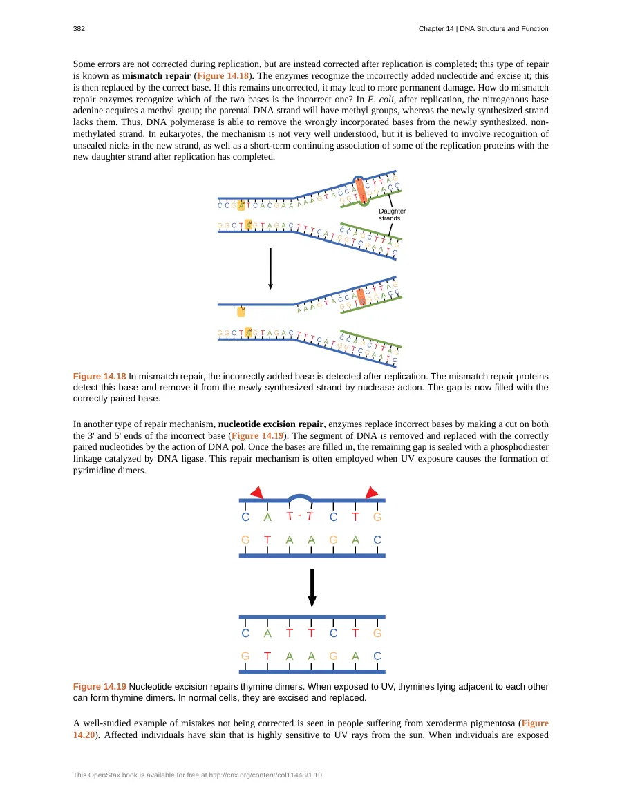
Mismatch Repair Mechanism
1 / 5How to study biology
Biology asks two things of you: how a process works, and how we know. These habits serve both, and each is shown on Chapter 14. They need only a pen and paper; where Hemle saves you time, we point to what it made.
Learn each experiment as question, method and result
Classic experiments come up in exams because they test reasoning: given the setup, predict what was found. For each one, write the question it asked, what was done, what was found, and which idea it ruled out.
In Chapter 14
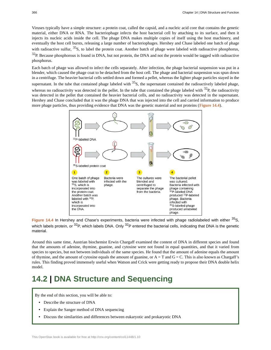
The chapter is built on them: Griffith; Avery, MacLeod and McCarty; Hershey and Chase; Chargaff (pp. 2–4); and Meselson and Stahl (pp. 12–13).
Hemle’s quiz on Hershey and ChaseDraw the process, then give each part its job
A mechanism is easier to recall as a picture than as a list. Draw it from memory, label every part with what it does, then compare your drawing with the book’s.
In Chapter 14
At the replication fork: helicase, single-strand binding proteins, topoisomerase, primase, DNA polymerase III and I, and ligase, each with one job. Table 14.1 lists them (p. 16).
Hemle’s cards on the replication forkAsk why it has to happen that way
Many biology facts follow from one constraint. Find the constraint and the facts stop being separate things to memorize.
In Chapter 14
The lagging strand is made in short Okazaki fragments because DNA polymerase can only extend a strand in the 5′ to 3′ direction, and the two strands of the helix run in opposite directions (p. 15).
Hemle’s answer on the two strandsTest yourself with problems, not rereading
Close the book and work problems: predict a result, fill in a ratio, explain an outcome. Getting one wrong shows you what to reread.
In one study, college students who spent their study time recalling a science text did better a week later, on questions about its facts and on questions that asked them to reason from it, than students who spent the same time drawing a concept map with the text open. (Karpicke and Blunt, 2011)
In Chapter 14
Chargaff’s rules make quick ones. If a DNA sample is 20% adenine, how much is guanine? (30%: thymine matches adenine, and the remaining 60% splits evenly between guanine and cytosine. p. 4)
Hemle’s cards on Chargaff’s rulesSet look-alikes side by side
Biology is full of pairs that differ in one way that matters. Put each pair in two columns and write the difference in a sentence.
In Chapter 14
Prokaryotic and eukaryotic replication (Table 14.2, p. 17), proofreading during replication and mismatch repair after it (pp. 19–20), transitions and transversions (p. 21).
Hemle’s cards on mismatch repairEvery card on three more concepts
Flashcards Hemle wrote on the chapter, each set whole: the question, the answer behind it, and the pages it cites. Try each one before you open it.
Chargaff's Rules
4 cards from pages 4–5
What are Chargaff's rules regarding the composition of DNA?Show answerHide answer
Chargaff's rules state that the amount of adenine equals the amount of thymine (A = T), and the amount of cytosine equals the amount of guanine (G = C). These quantities vary between species but remain constant among individuals of the same species.
True or False: In all DNA molecules, the concentration of adenine, thymine, guanine, and cytosine are equal.Show answerHide answer
False. Chargaff's rules demonstrate that while A = T and G = C, the four bases are not found in equal quantities overall, and their relative amounts vary from species to species.
If a DNA sample contains 20% adenine, what is the percentage of thymine, guanine, and cytosine according to Chargaff's rules?Show answerHide answer
Following Chargaff's rules, if adenine is 20%, then thymine must also be 20% (A = T). This leaves 60% for the remaining bases, which must be split equally between guanine and cytosine (G = C), resulting in 30% guanine and 30% cytosine.
Chargaff's finding that A = T and G = C was essential for the development of the ________ model of DNA.Show answerHide answer
double helix
Meselson-Stahl Experimental Evidence
5 cards from page 13
What observation after one generation of growth in ¹⁴N media led Meselson and Stahl to conclude that the conservative model of DNA replication was incorrect?Show answerHide answer
After one generation, the DNA formed a single band at an intermediate density position between the ¹⁵N and ¹⁴N levels. This result contradicts the conservative model, which would have predicted two distinct bands representing purely "heavy" and purely "light" DNA.
Why was a second generation of growth in ¹⁴N necessary for Meselson and Stahl to rule out the dispersive model?Show answerHide answer
While the intermediate band after one generation was consistent with both semi-conservative and dispersive models, the second generation produced two distinct bands: one at the intermediate position and one at the ¹⁴N position. This pattern is only consistent with semi-conservative replication, whereas the dispersive model would have predicted a single band of decreasing density.
In the Meselson-Stahl experiment, why does DNA grown in ¹⁵N sediment to a lower level in a cesium chloride gradient than DNA grown in ¹⁴N?Show answerHide answer
The ¹⁵N isotope is "heavy," making the DNA molecules that incorporate it denser than those containing only the lighter ¹⁴N isotope. During density gradient centrifugation, molecules band according to their density, with denser molecules moving to a lower position in the gradient.
A student claims that the intermediate density band observed after the first generation of the Meselson-Stahl experiment proves the dispersive model. Why is this incorrect?Show answerHide answer
This is a common misconception because the intermediate band is consistent with both the semi-conservative and dispersive models. The intermediate band only indicates that each DNA molecule contains a mix of "old" and "new" material, but it does not distinguish between the two models until a second generation of replication is observed.
In the Meselson-Stahl experiment, the observation of two distinct bands after two generations of replication in ¹⁴N media provides evidence for the __________ model of DNA replication.Show answerHide answer
semi-conservative
Asymmetric Replication Fork Dynamics
4 cards from page 15
Why must the lagging strand be synthesized in discontinuous fragments during DNA replication?Show answerHide answer
Because the DNA double helix is anti-parallel and DNA polymerase can only extend a new strand in the 5' to 3' direction, the strand oriented 3' to 5' relative to the replication fork must be synthesized in short, discontinuous stretches known as Okazaki fragments.
True or False: DNA polymerase can synthesize both the leading and lagging strands continuously because it is a highly efficient enzyme.Show answerHide answer
False. DNA polymerase can only synthesize in the 5' to 3' direction. Due to the anti-parallel nature of the DNA double helix, the lagging strand must be synthesized in discontinuous Okazaki fragments.
In a hypothetical scenario where a cell strain has a mutation that prevents the joining of Okazaki fragments, which enzyme is most likely defective?Show answerHide answer
DNA ligase, as it is the enzyme responsible for sealing the gaps between Okazaki fragments by catalyzing the formation of phosphodiester linkages.
The lagging strand is synthesized in short stretches called ___________, which require a new RNA primer for each segment.Show answerHide answer
Okazaki fragments
Made in Hemle on 28 October 2026: every card on 3 concepts, word for word, of the 5 sets we asked for (another is in the window above). The set on “Enzymatic Roles in Replication” isn’t shown: one of its cards came back with “{cloze}” where its blank should be. From Chapter 14 of Biology, first edition (2013), by OpenStax, under a CC BY 4.0 licence. Download for free at openstax.org/details/books/biology. The cards, quiz and answers were made by Hemle’s AI; OpenStax doesn’t endorse Hemle.
A whole quiz on Hershey and Chase
Hemle’s five questions on Hershey-Chase Isotopic Labeling, as they came: two on why each label works, two that ask what the experiment found, and one that corrects a student’s claim. Each answer quotes the chapter’s sentence it rests on.
Question 1 of 5
In the Hershey-Chase experiments, why was ³⁵S used specifically to label the viral protein coat rather than nucleic acids?
Show answerHide answer
Answer C: Sulfur is present in proteins but is absent from DNA.
Page 4 explains that sulfur is found in protein but not in nucleic acids such as DNA. Radioactive sulfur (³⁵S) therefore selectively tags the viral protein coat. Ask yourself: Which chemical element is distinct to proteins compared to nucleic acids in viral structures?
“Hershey and Chase labeled one batch of phage with radioactive sulfur, 35S, to label the protein coat.”
Question 2 of 5
Which statement correctly describes why Alfred Hershey and Martha Chase used radioactive ³²P in their bacteriophage cultures?
Show answerHide answer
Answer A: Phosphorus is an integral element of DNA but is not found in protein.
As stated on page 4, phosphorus is found in DNA but not in protein. This chemical distinction allowed Hershey and Chase to selectively tag phage DNA with radioactive phosphorus. Ask yourself: What molecular component of bacteriophages contains phosphorus atoms?
“Because phosphorous is found in DNA, but not protein, the DNA and not the protein would be tagged with radioactive phosphorus.”
Question 3 of 5
A researcher repeats the Hershey-Chase protocol using bacteriophages labeled with ³⁵S. After allowing bacterial infection, agitating the mixture in a blender, and centrifuging the suspension, where will the majority of the radioactivity be detected?
Show answerHide answer
Answer A: In the supernatant containing the detached viral coats
According to page 4, the blender detaches phage coats from the bacterial cell surface, and centrifugation pellets the heavier bacterial cells. In the ³⁵S batch, the empty viral protein coats remain in the lighter supernatant where the radioactivity is detected. Ask yourself: Where do the detached, lighter viral protein coats remain after centrifugation?
“In the tube that contained phage labeled with 35S, the supernatant contained the radioactively labeled phage, whereas no radioactivity was detected in the pellet.”
Question 4 of 5
How did the post-centrifugation distribution of ³²P differ from that of ³⁵S in the Hershey-Chase experiment?
Show answerHide answer
Answer A: ³²P was detected in the bacterial pellet, whereas ³⁵S was found in the supernatant.
Page 4 highlights that in the tube with ³²P-labeled phage, radioactivity was detected in the pellet containing the heavier bacterial cells. Conversely, in the tube with ³⁵S, radioactivity was present only in the supernatant with no radioactivity detected in the pellet. Ask yourself: Which isotope was carried into the heavy bacterial pellet at the bottom of the centrifuge tube?
“In the tube that contained the phage labeled with 32P, the radioactivity was detected in the pellet that contained the heavier bacterial cells, and no radioactivity was detected in the supernatant.”
Question 5 of 5
A student claims: 'In the Hershey-Chase experiment, both the protein capsid and the viral DNA enter the bacterial host, but only DNA directs replication.' What is incorrect about this statement?
Show answerHide answer
Answer B: The protein capsid does not enter the host cell; it remains attached outside and is detached.
Page 4 notes that bacteriophages attach to the cell surface and inject only their nucleic acids inside the cell. The empty protein coats remain outside and can be sheared off in a blender, leaving the supernatant radioactive in ³⁵S trials. Ask yourself: Does any part of the bacteriophage protein coat actually enter the bacterial cytoplasm?
“The bacteriophage infects the host bacterial cell by attaching to its surface, and then it injects its nucleic acids inside the cell.”
Made in Hemle on 28 October 2026: the whole quiz, word for word. Question 1’s explanation says page 4 explains that sulfur is in protein but not in nucleic acids; page 4 says only that sulfur labelled the protein coat. From Chapter 14 of Biology, first edition (2013), by OpenStax, under a CC BY 4.0 licence. Download for free at openstax.org/details/books/biology. The cards, quiz and answers were made by Hemle’s AI; OpenStax doesn’t endorse Hemle.
One more question, word for word
The leading and lagging strands are the chapter’s most often confused pair, so we asked Chat for the difference. Here is Hemle’s answer in full, with the pages it cites in brackets.
What's the difference between the leading strand and the lagging strand?
Show Hemle’s answerHide Hemle’s answer
The leading strand is made continuously toward the replication fork, while the lagging strand is made away from the fork in short pieces called Okazaki fragments. Both differences arise because DNA polymerase can extend DNA only in the 5′ to 3′ direction, while the two parental DNA strands run in opposite directions (p. 15).
Feature Leading strand Lagging strand Direction relative to the replication fork Synthesized toward the fork Synthesized away from the fork Pattern of synthesis Continuous In short Okazaki fragments Primers needed One primer can start its synthesis A new primer is needed for each fragment At the origin of replication, helicase separates the DNA strands and forms a replication fork. Because the parental strands are antiparallel, DNA polymerase can follow one template toward the fork while extending the new strand 5′ to 3′. That is the leading strand. On the other template, synthesis must proceed away from the fork in separate stretches, producing the lagging strand (pp. 15, 24).
The distinction is useful because it explains why replication proceeds differently on the two sides of the fork and why the lagging strand needs extra steps. The RNA primers are replaced with DNA, and DNA ligase seals the gaps between Okazaki fragments to make a continuous strand (p. 15). Figure 14.14 illustrates this arrangement: helicase opens the DNA, DNA polymerase makes the leading strand continuously and the lagging strand in fragments, and ligase joins those fragments (p. 15).
Asked in Hemle’s Chat on 28 October 2026, each question in a new chat; the answer is word for word. One of the questions we asked isn’t shown: “How did Meselson and Stahl show that DNA replication is semi-conservative?” Its answer missed information that’s on page 13: the two bands after the second generation. From Chapter 14 of Biology, first edition (2013), by OpenStax, under a CC BY 4.0 licence. Download for free at openstax.org/details/books/biology. The cards, quiz and answers were made by Hemle’s AI; OpenStax doesn’t endorse Hemle.
Common questions
What’s the best way to study biology?
Learn each experiment as its question, method and result; draw processes such as DNA replication from memory and give each part its job; ask why each step has to happen the way it does; test yourself with problems instead of rereading; and set look-alike terms side by side. This page shows each on a real chapter on DNA.
Can AI help me study biology?
Yes: AI can turn your chapter into flashcards and quizzes, answer questions about it, and bring each concept back for review. Hemle works from the chapter you upload, so the terms and examples are your book’s, and every card, question and answer names its page.
Can Hemle get things wrong?
Yes. Answers are written by AI and can be wrong, which is why each one names its pages. Of the two questions we asked Chat about this chapter, one answer missed information that’s on page 13, so it isn’t shown here. When an answer and your book disagree, trust the book.
Does it work with my biology textbook?
Yes, if you can upload the chapter: a PDF, Word or PowerPoint file up to 20 MB. Your free chapter can be up to 100 pages. Hemle maps the chapter’s concepts, then writes cards, quizzes and answers from it.
Which edition is the chapter on this page from?
The first edition of OpenStax’s Biology (2013), published under a CC BY 4.0 licence. OpenStax has since published a second edition, so your course’s book may differ. Hemle works from whatever chapter you upload.
Is Hemle free?
Your first chapter is free, and you don’t need a card to start. Paid plans lift the limits when you want more.
Is my file private?
Your file stays in your own library, and it isn’t used to train AI. You can delete it, or your whole account, at any time.
About the chapter
Chapter 14, “DNA Structure and Function”, of Biology, first edition, by Connie Rye, Robert Wise, Vladimir Jurukovski, Jean DeSaix, Jung Choi and Yael Avissar. Published by OpenStax, Rice University, in 2013; ©2017 Rice University. Textbook content produced by OpenStax is licensed under a Creative Commons Attribution 4.0 International licence (CC BY 4.0). Download for free at https://openstax.org/details/books/biology.
What we changed: we used this chapter alone, pages 375 to 400 of OpenStax’s PDF of the first edition (279 MB), and uploaded those 26 pages as they are. Page numbers on this page are that file’s, as Hemle gives them: its page 1 is the book’s page 363. The page images are rendered from it, unchanged. The flashcards, quiz and answers were written by Hemle’s AI.
The address above now opens the book’s second edition, which OpenStax publishes under a different licence. This page uses the first edition only. OpenStax and Rice University don’t endorse Hemle or this page.
More from Hemle
Anatomy and physiology
Cards, a quiz and answers from a real chapter on the heart, with how to study A&P.
Upload a textbook to AI
Ask your chapter anything; every answer names the pages it comes from.
PDF to flashcards
Flashcards from your chapter, each naming the page it came from.
AI quiz generator
Multiple-choice questions on your chapter, with an explanation for every answer.
The AI tutor
A real session on another chapter: Hemle teaches it one concept at a time and cites the page as it goes.